
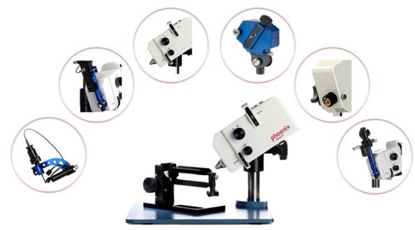
Instrument name | Phoenix Micron IV Retinal Imaging Microscope |
Manufacturer | Phoenix Research Lab |
Type number | Phoenix Micron Ⅳ |
Specifications | 1.Imaging Modes: Bright-field imaging, angiography, fluorescence imaging 2.Retinal Imaging Resolution: 3 µm (mouse); Retinal Imaging Pixel Size: 2 µm (mouse) 3.Dynamic Imaging Frame Rate: 24 fps to 0.5 fps; Dynamic Range: 60 dB; Field of View: ≥ 50° 4.Image Sensor: Custom 3-chip CCD for enhanced sensitivity and signal-to-noise ratio (SNR) 5.Data Output: Supports image and video output; Focus Range: from the lens to the retinal surface, depth of focus: 20 µm 6.4-position filter wheel, dual-compensated lamp and filter configuration for greater flexibility, supporting bright-field and fluorescence imaging modes 7.Experimental Stage: Capable of 3D tilting and rotation to adjust the eye angle of mice and rats for clear imaging |
Function and applications | The Micron Ⅳ Retinal Microscopy Imaging System delivers high-resolution retinal images with a resolution of 3 µm. It supports bright-field and fluorescence imaging (Ex. CFP, GFP, mCherry, etc.), enabling direct switching from bright-field to fluorescence imaging. The Image-Guided Laser System features user-friendly design, allowing operators to precisely and reliably control the position, size and intensity of the delivered laser to meet diverse research requirements.Beyond retinal imaging, the system can be further expanded with multiple functions including laser injection guidance and laser module, high-resolution OCT, slit lamp imaging, ERG electrophysiology, etc., providing a comprehensive solution for ophthalmic research. |
Contacts:Xuejiao Han | |
Contact number: 65880320 | |
E-mail: xjhan@suda.edu.cn | |
Editor: Xuejiao Han